The main work from my PhD is now out on bioRxiv (Ton et al. 2022) 😃.
In a collaboration with Mai-Linh Ton, Novo Nordisk, Göttgens, Benito-Guitierrez and Marioni labs, we used single-cell transcriptomics to molecularly profile early rabbit development.
Summary
What is Single-Cell Transcriptomics? 🧬
Single-cell transcriptomics is a method to univesally profile the genes that are expressed (‘turned on/off’) in individual cells of a tissue/organism. It allows you to characterise populations of cells in a biological sample at extremely high-resolution and study their molecular properties.
Why the rabbit? 🐇
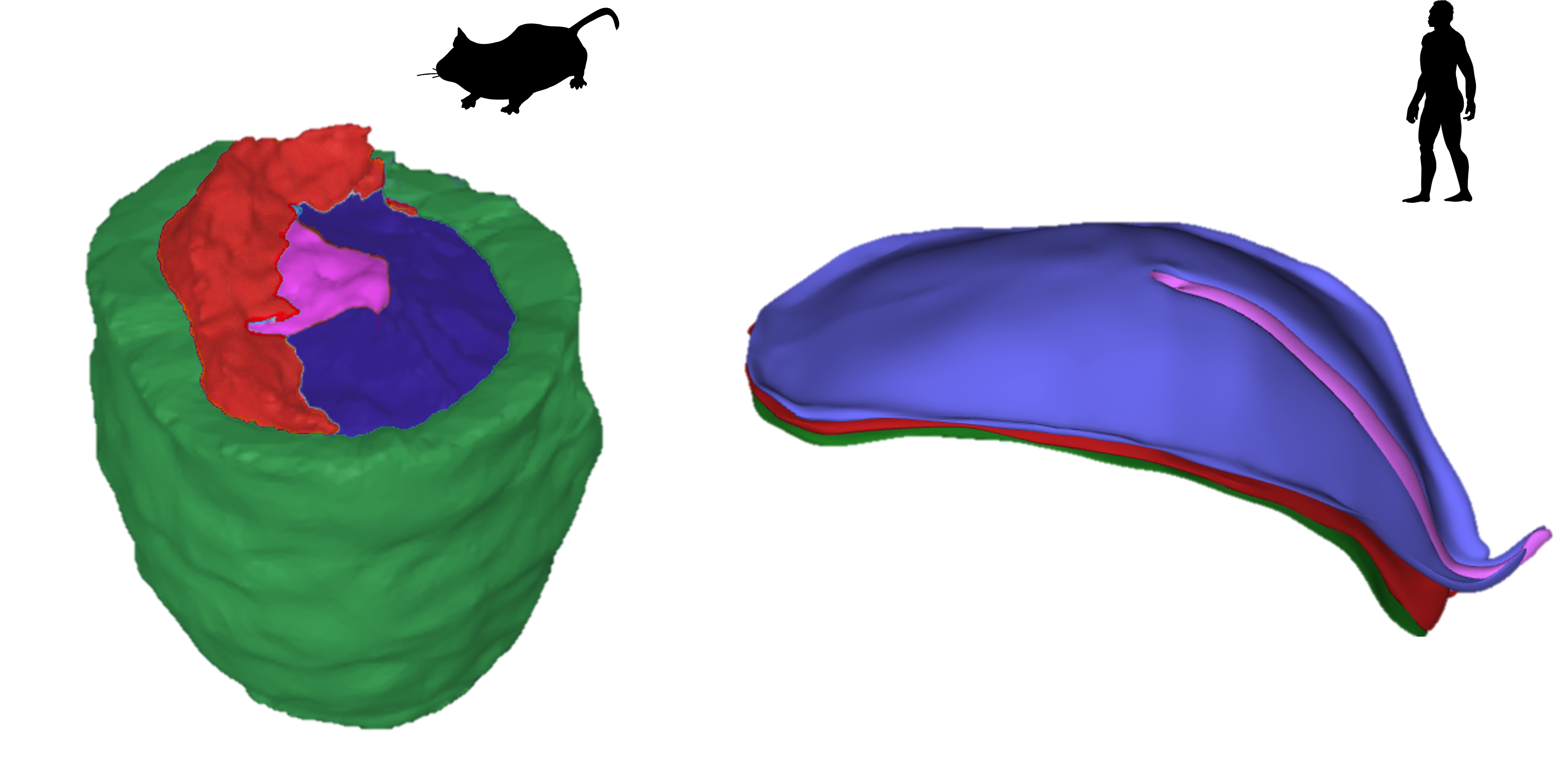
Due to ethical and technical challenges, our understanding of early human development is mostly derived from the mouse. However many aspects of early mouse development diverge from other mammals, complicating how we translate findings to humans. For example, the early mouse embryo has a cup-shape, while most mammals including rabbits and humans develop as a flat-disc.
What did we find? 🔍
- We used single-cell transcriptomics to molecularly profile 146,133 cells across days 7, 8 and 9 of rabbit development.
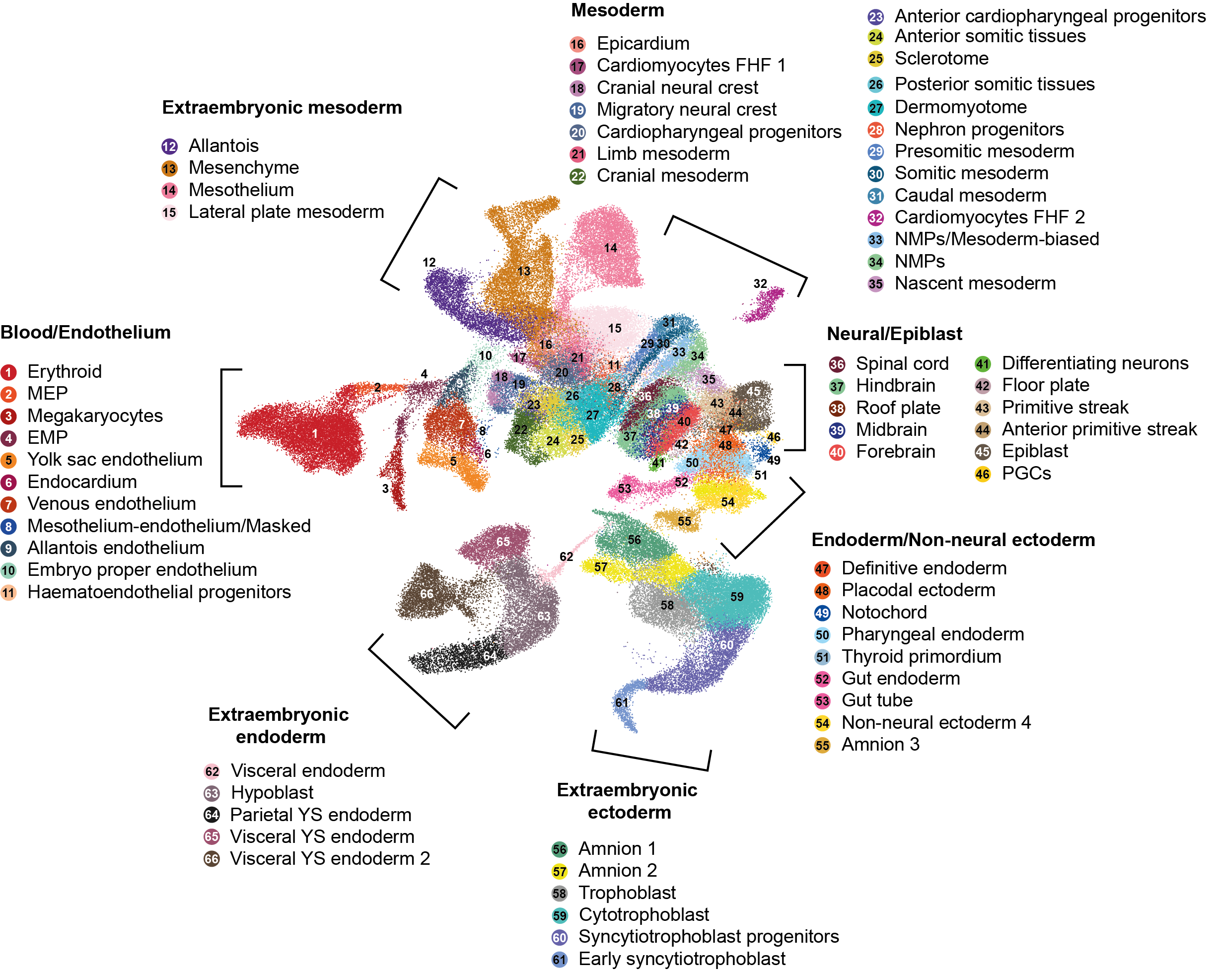
Figure 1: In this UMAP plot each point represents a cell, positioned in relation to the genes it expresses (e.g. cells closer together generally have more similar gene expression). We identify 66 different types of cells in our dataset.
- We compared the molecular changes that take place during rabbit and mouse development and find that certain types of cells differ more than others between the two species - particularly cells involved in the development of supportive ‘extra-embryonic’ structures1. This has implications for how we interpret results from specific tissues in mice and rabbits.
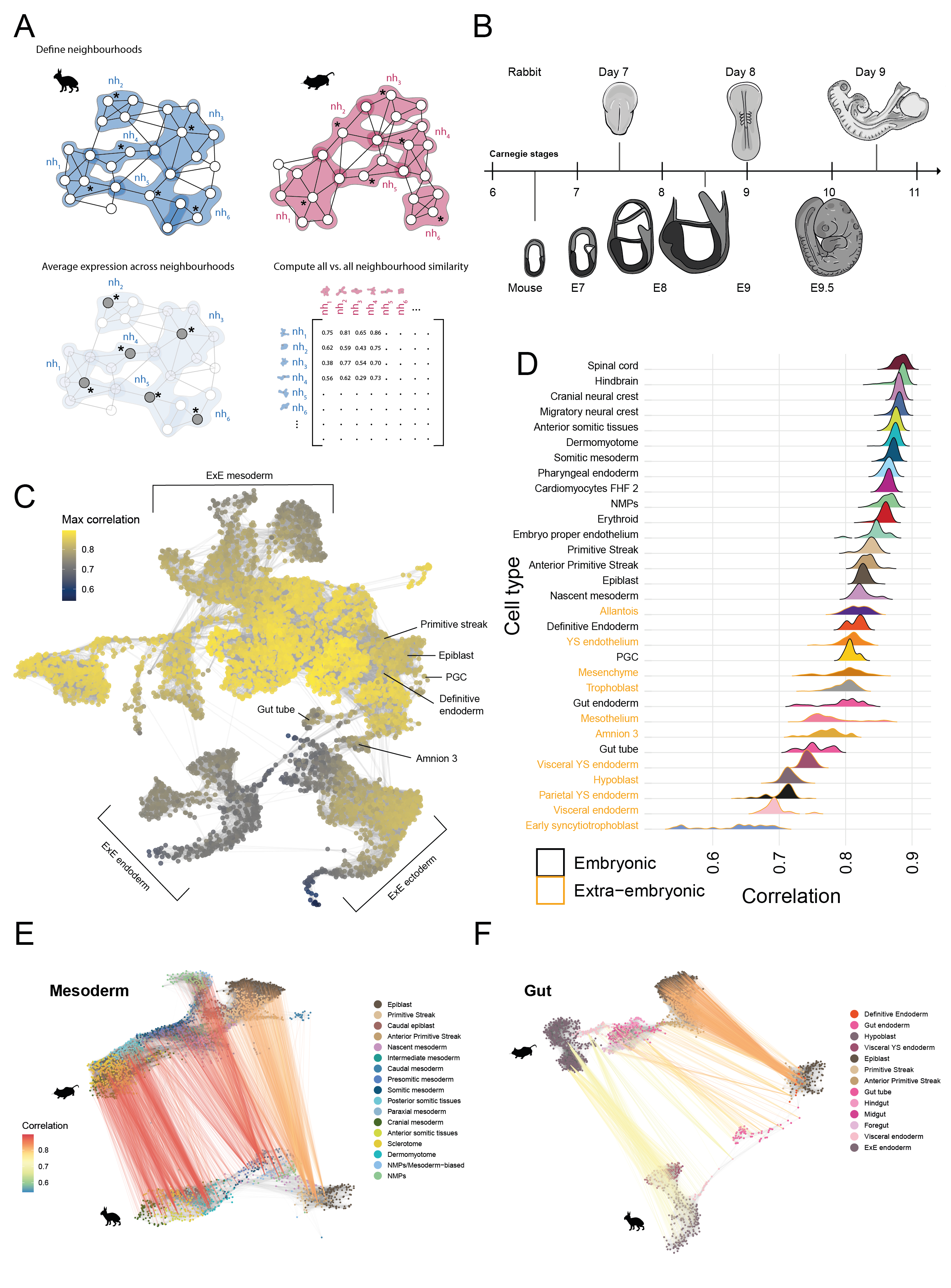
Figure 2: We developed a new approach to compare gene expression datasets from the rabbit and mouse (schematic in A). The ridgeline plot in D) shows that extra-embryonic cell types (orange) are the most dissimilar/least correlated between the two species. We also compared cells across the development of specific tissues, such as the gut (F).
- We also find that the rabbit is particularly suitable to study the process of implantation - which is difficult to study in mice/humans and which is important for understanding pregnancy-related disorders. We find similarities with mice but also differences that are in common with humans.
![Implantation is facilitated by trophoblast cell types, which are enriched in our dataset (A). In particular, we observe a trajectory of trophoblast differentiation (B) and identify genes which are expressed over the course of their development (C). In particular we find that DLX5 and DLX6 are expressed, which are not expressed in mice trophoblast cells. The is notable since DLX5/6 expression has been observed in human trophoblast cells and is implicated in pre-eclampsia [@Zadora2017-yo].](figs/fig2_trophoblast.png)
Figure 3: Implantation is facilitated by trophoblast cell types, which are enriched in our dataset (A). In particular, we observe a trajectory of trophoblast differentiation (B) and identify genes which are expressed over the course of their development (C). In particular we find that DLX5 and DLX6 are expressed, which are not expressed in mice trophoblast cells. The is notable since DLX5/6 expression has been observed in human trophoblast cells and is implicated in pre-eclampsia (Zadora et al. 2017).
- Moreover, we show that by using large molecular datasets from the rabbit and mouse, we are able to gain greater insight into the very limited datasets that exist from humans and non-human primates. We are able to identify very specific types of cells in primate datasets with very few cells.
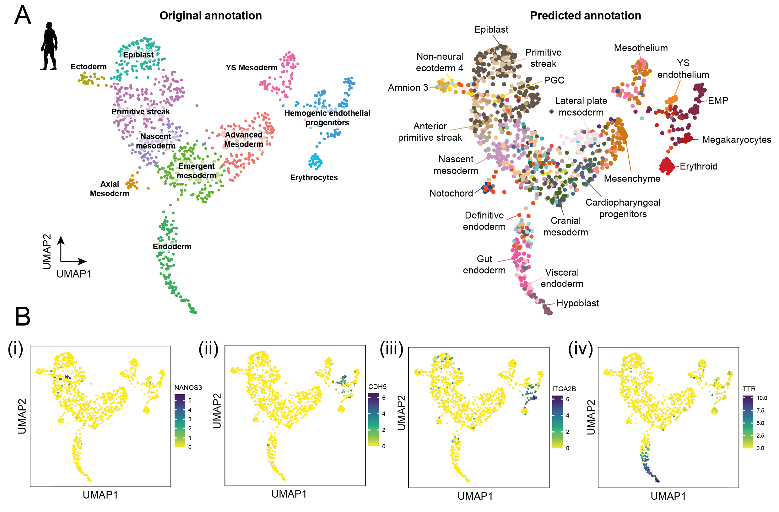
Figure 4: We used our rabbit dataset to train a computational model which can be used predict the identity of cells from a human dataset (A). Our predictions are consistent with the expression of genes known to be specific to particular cell types (B). For example CDH5 is a known marker of endothelium (Bii).
- Finally, we took advantage of the large size of the rabbit embryo to study blood development. We find that a particular cell type called the mesothelium, plays a more important role in signalling the formation of blood vessels than perhaps previously recognised
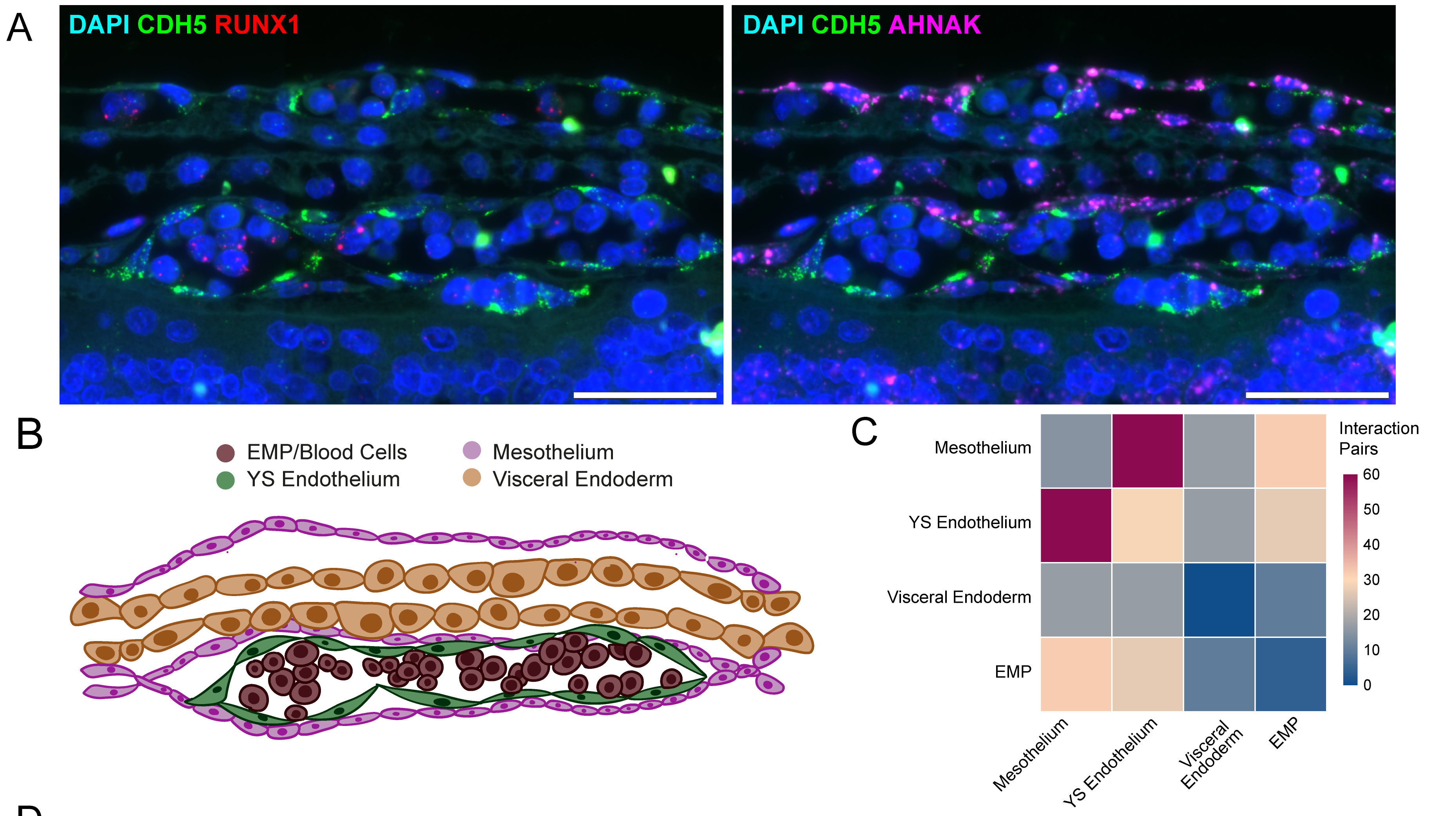
Figure 5: We show that the mesothelium is in direct contact with the endothelium (A,B), which is invovled in blood vessel formation. From our gene expression data we also predict a large number of molecular interactions between these cell types (C) .
Summary ✨
In summary, our work demonstrates how comparing across species can help to decipher early human development and potentially improve the translation of findings from model organisms.
To find out more, you can read on below or the tweetorial here.
Alternatively, you can read the full preprint on bioRxiv: https://doi.org/10.1101/2022.10.06.510971.
Termed extra-embryonic as they are not directly involved in the development of the embryo itself. Examples include the allantois, the precursor to the umbilical cord. ↩︎